The uracil-DNA glycosylase UNG protects the fitness of normal and cancer B cells expressing AID

Safavi S, Larouche A, Zahn A, Patenaude AM, Domanska D, Dionne K, Rognes T, Dingler F, Kang SK, Liu Y, Johnson N, Hébert J, Verdun RE, Rada CA, Vega F, Nilsen H, Di Noia JM. The uracil-DNA glycosylase UNG protects the fitness of normal and cancer B cells expressing AID. NAR Cancer. 2020 Aug 27;2(3):zcaa019. doi: 10.1093/narcan/zcaa019. Erratum in: NAR Cancer. 2021 Jan 06;3(1):zcaa045. PMID: 33554121; PMCID: PMC7848951.

Studies of human gut plasma cells: implications for vaccination and treatment of chronic inflammation


Studies of human gut plasma cells: implications for vaccination and treatment of chronic inflammation

Morbidity and mortality caused by infections in the gastrointe¬stinal tract is a major health problem worldwide. Gut infections are responsible for approxi¬mately 10 million deaths annually of children younger than 5 years of age. For the majority of these diseases, there are no available effective vaccines. The gut is the habitat for trillions of commensal microbes (microbiota) that are beneficial for the host. However, disruption to the normal balance between the gut microbiota and the host (termed dysbiosis) has been associated with inflammatory bowel disease, obesity, allergy and asthma, autoimmune diseases and neurological disorders. The microbiota has thus become a very attractive target for therapeutic interventions. Plasma cells (PCs) are one of the most prominent immune cell-types in the gastrointestinal tract. They produce secretory antibodies (mainly immunoglobulin A (IgA)) that are actively transported into the gut lumen where they protect against invading enteropathogens. Moreover, we and others have shown that secretory antibodies reinforce homeostasis in the gut by regulating the composition of the microbiota.
The prevailing scientific dogma states that PCs of the gut have short half-lives. However, we have provided conclusive evidence that intestinal PCs per¬sist for decades in humans. This finding fundamentally changes the concept of gut humoral immunity with implications both for vaccine development and for therapeutic strategies to target the microbiota. This project directly builds on this paradigm-shifting discovery and our main objectives are to: 1) Identify molecular mechanisms underlying the selection of long-lived intestinal PCs; 2) Identify and characterize members of the microbiota that are targeted by long-lived PCs.
We believe results from this project will have far-reaching clinical implications: 1) Understanding the molecular mechanisms that drive the selection of long-lived PCs is of great importance for the design of vaccines with long-lasting protective effects. 2) There is good reason to believe that commensals that induce persistent IgA responses are particularly important for gut homeostasis and that they therefore would be very attractive targets for therapeutic interventions in diseases associated with dysbiosis.

Distribution of plasma cells (CD38, red) and CD45+ cells (green) in the small intestine

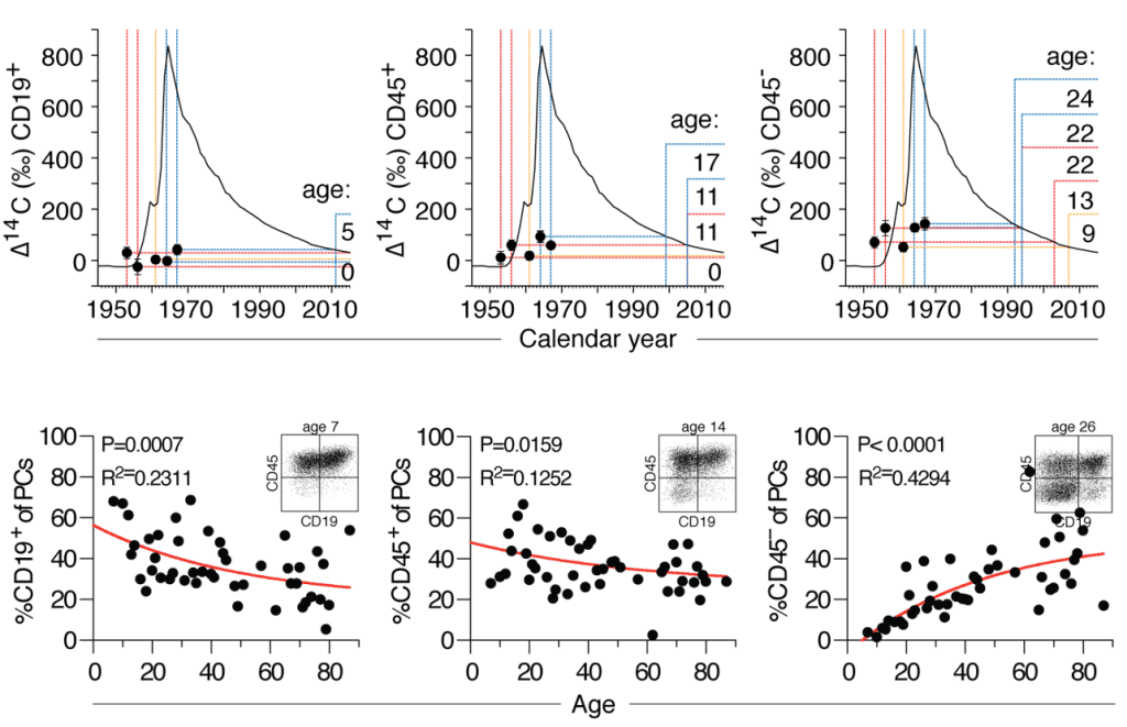
Age of plasma cell subsets (CD19+, CD45+, CD45-) derived from small intestine of 5 individuals based on carbon-14 concentrations in genomic DNA (upper). Percentage of plasma cell subsets changes with age (lower). For details see Landsverk et al. J Exp Med, 2017.

(A) The frequency of IGHV gene segments used by plasma cells. (B) The level of somatic hypermutation among plasma cells. (C) Morisita-Horn indices for clonal similarity between PC subsets. (D) Clonally related sequences assigned between plasma cell subsets illustrated in a Circos plot.

Dissecting differentiation and diversification of gut macrophages


Dissecting differentiation and diversification of gut macrophages

Macrophages reside in virtually all organs and play a key role in host defense and tissue homeostasis. Tissue macrophages are particularly important in the gut where they defend the body against a wide variety of pathogens and toxic substances.
However, gut macrophages also contribute to pathology. Aberrant activation of macrophages plays an important role in disorders such as inflammatory bowel disease (IBD; ulcerative colitis and Crohn’s disease), necrotizing enterocolitis in infants and mobility dysfunctions. Importantly also, tumor-associated macrophages in colorectal cancer can promote cancer growth.
Although gut macrophages are instrumental in both host defense and pathology, knowledge about their origin, heterogeneity and functional properties is scarce. To increase our understanding of macrophages in the intestine we recently performed transcriptional, phenotypical and functional profiling of tissue macrophages in the human small intestine. We found that macrophages consist of several transcriptional states and by examining gut macrophages in a unique organ transplantation setting we found that blood monocytes emigrated into duodenal transplants and differentiated, through short-lived intermediates, into mature macrophages that survive for months.
To get a deeper understanding of macrophage diversity and functions we have applied single cell RNA sequencing to analyze macrophages isolated from the intestinal mucosa and the underlying smooth muscle layers (muscularis propria). We find that there is an unexpected macrophage heterogeneity within both compartments and between the compartments. Our findings raise several questions we will address in this project: What are the microenvironmental signals that imprint macrophage identity and diversity? Since macrophages are so diverse, is it possible to reprogram macrophages? And do they all originate from the same precursor cells?
We will apply an integrated approach combining high throughput single-cell technologies with unique clinical material and 3D organ cultures. Computational integration of the data will provide a spatiotemporal reconstruction of how monocyte-to-macrophage differentiation and diversification in the human gut is regulated and reveal functional properties.

Immunofluorescence image of macrophages (CD68/CD163, green) in mucosa and submucosa of colon. Lyve1+ macrophages (yellow) are only found in submucosa.
UMAP clustering (A), dot plot (B) and UMAP (C) of selected genes, and pseudotime trajectory (D) of macrophages derived from colonic mucosa
Spatial Trancriptomics visualizing S100A8/A9+ (left) and HLA class II high macrophages (right) in colon.

Understanding the development of resident memory T cells in the human small intestine using integrative multiomic approaches


Understanding the development of resident memory T cells in the human small intestine using integrative multiomic approaches (PI Raquel Bartolome Casado)

Tissue-resident memory T cells (TRM) are permanently lodged in barrier tissues like the gut, and protect the host against pathogens. However, the biological pathways that enable the long-term survival of TRM are poorly understood. By exploiting a unique human intestinal transplantation setting where we can directly distinguish persisting TRM from circulating T cells, we have recently shown that the human small intestine contains large populations of long-lived CD4+ and CD8+ T cells. We will now use this transplantation model to identify mechanisms and anatomical niches that promote and support the development and maintenance of TRM cells. To this end, we will partner with Sarah Teichmann at the Wellcome Sanger Institute in Cambridge, UK, who is a co-founder of the Human Cell Atlas Consortium and a world-leading expert in the field of single-cell genomics and its application to T-cell biology. Postdoc Raquel Bartolome-Casado will spend two years (from June-2021) in Dr. Teichmann’s lab to apply cutting-edge multiomic single-cell technologies, including parallel epigenetic, transcriptomic and T cell receptor analysis, to decipher the developmental pathways of human gut TRM cells. This project will provide unprecedented knowledge on the mechanisms of immune memory development in the human gut, which has major implications for long-lasting oral vaccination strategies and treatments of intestinal immune disorders involving persistent pathogenic T cells.

Drawing of transplanted pancreas with a piece of duodenum (left). Percentage of donor CD8+ T  cells  in donor duodenum at different time points after transplantation (right). CD103- lamina propria cells (LPCs, red), CD103+ LPCs (blue), and CD103+ intraepithelial cells (IELs) (green). Adapted from Casado et al. JEM, 2019.

Identifying novel pathways to treat Hirschsprung-associated enterocolitis


Identifying novel pathways to treat Hirschsprung-associated enterocolitis

Hirschsprung’s disease (HD) is a common cause of neonatal bowel obstruction. It is characterised by an absence of enteric neurons (aganglionosis) in the distal bowel. The aganglionosis involves the internal anal sphincter and extends orally, most often to the sigmoid colon. The aganglionic segment of the colon fails to relax, causing a functional, severe bowel obstruction. Current treatment is surgical resection of aganglionic bowel. Unfortunately, many children have problems after surgery, particularly in the first years of life. Postoperative problems include constipation, obstructive defecation, fecal incontinence and Hirschsprung-associated enterocolitis (HAEC).
HAEC is a dangerous clinical symptom complex characterized by abdominal distension, fever and diarrhoea. In addition, HAEC has been shown to be associated with reduced functional outcome, particularly fecal incontinence. HAEC occurs in 30–60% of HD patients and may occur both pre-and postoperatively. In some children, HAEC is the presenting symptom of HD, whereas others have one or more episodes after surgery. HAEC is most frequent the first year postoperatively, but some children continue to have relapsing HAEC episodes for many years. HAEC aetiology is poorly understood. Interestingly, this symptom complex does not occur in otherwise healthy children with severe chronic constipation or other bowel disorders. The aim of this project is to increase current understanding of HAEC aetiology with the goal to improve treatment for this condition, which causes severe morbidity and even may be life-threatening.

We hypothesize that the lack of enteric neurons affects the development and function of immune cells in the gut and that disturbances of mucosal immunity in patients with HD is an underlying cause for HAEC.
The aim of this project is to study the mucosal immune system in the gut of HD patients by computational integration of single cell RNA sequencing, Spatial Transcriptomics and high dimensional immune-imaging of colonic tissue from HD patients and age-matched controls.

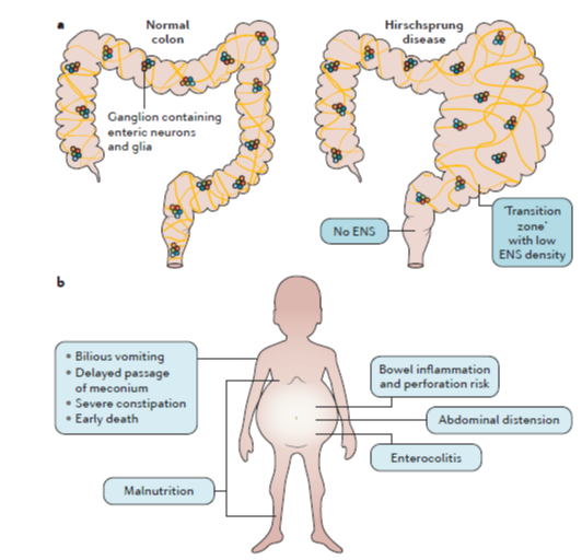
Hirschsprung disease. a) In children with Hirschsprung disease, all enteric neurons and associated glia are absent from the distal aganglionic bowel (right). The aganglionic segment of the colon fails to relax, causing a functional, severe bowel obstruction. b) Gives an overview of symptoms of Hirschsprung (Adapted from (Heuckeroth, 2018).
UMAP of all T cells derived from colon of children < 2 years

Identifying biomarkers to predict graft versus host disease in hematopoietic stem cell transplanted patients


Identifying biomarkers to predict graft versus host disease in hematopoietic stem cell transplanted patients

30-50% of allo-HSCT patients suffer from graft versus host disease (GVHD); a disease with very high mortality rate. Biomarkers that identify patients at risk for GVHD is important to treat these patients before or early in progression of disease. We have collected blood samples at multiple timepoints from more than 100 patients after transplantation. We are currently examining these samples by two complementary approaches: 1) T cells are assessed with CYTOF technology that are able to phenotype the cells simultaneously with 40 antibodies. 2) Plasma proteins are measured with a commercially available kit from Olink. This allows us to quantitate 1536 proteins in every sample. Comparing the phenotype of T cells and levels of proteins over time in patients that develop GVHD with those that do not develop GVHD, our goal is to identify clinical relevant biomarkers.

Heatmap of 1536 proteins expressed in plasma of patients with and without acute gut GVHD

Spatial transcriptomics to predict treatment response in Crohn’s disease


Spatial transcriptomics to predict treatment response in Crohn’s disease (PI Espen Bækkevold)

Anti-TNF therapy is currently the first treatment choice for newly diagnosed patients with Crohn’s disease (CD). However, approximately 30% of CD patients have no effect of anti-TNF therapy, and because there are no clinical criteria to identify non-responders, all patients are initially given such treatment. The aim of this project is to identify biomarkers that with high accuracy predict which patients that will fail anti-TNF treatment. Clinical implementation of such biomarkers will personalize the treatment of CD with significant health benefits.
To untangle the cellular networks associated with durable remission upon anti-TNF therapy, we will take advantage of the most recent advances in experimental and computational methods and study the disease process in CD-lesions with two complementary high dimensional techniques:
1) Analyses of datasets from single-cell RNA sequencing (scRNAseq) of tissue-derived cells from inflamed and health intestine will give an unbiased characterization of the gene expression levels of all cells, and 2) the Spatial Transcriptomics (ST) method will reveal the spatial pattern of gene expression levels within the tissue.
Advanced bioinformatics to integrate the two methods will give unbiased and comprehensive tissue maps with unprecedented molecular resolution.
Based on this analysis we will construct a single cell transcriptome atlas with spatial information across tissues from both anti-TNF responders and non-responders. This will give a unique possibility to identify differences at the cellular and molecular level in time and space in the search for predictive biomarkers for treatment response.

Flow cytometry analysis of macrophages (macs) isolated from Crohn’s disease lesions (obtained by colonoscopy, right image) and adjacent uninflamed mucosa showing a large expansion of inflammatory macrophages in the lesions.

Naveen Parmar (Postdoc)

I was employed as a postdoctoral fellow and a researcher at CEMIR, NTNU Trondheim for 3, 5 years. I investigated the role of the epigenetic enzyme during gut homeostasis and infection-induced colitis in mouse models, so I have expertise in establishing mouse enteroids and colonoids. Usually, I used immunofluorescence and confocal microscopy methods to dissect new aspects of intestinal biology. Therefore, I am very excited to start my new project at Rikshospitalet, UiO, on the human gut system.